ETV Bharat / state

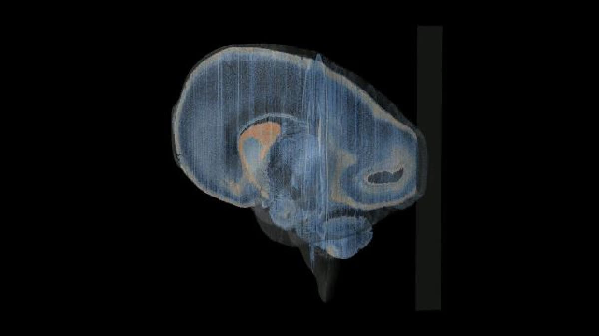
IIT Madras Releases World's First 3D Images Of Human Foetal Brain

This research is critical for India as the country accounts for nearly one-fifth of the world’s childbirths at 25 million each year.

Courtesy: IIT-Madras
The 3D image. (IIT-Madras Releases World's First 3D Images Of Human Foetal Brain)
author img

By ETV Bharat English Team

Published : Dec 11, 2024, 10:04 AM IST

Updated : Dec 11, 2024, 1:32 PM IST

Chennai: In a landmark feat, IIT Madras has released the world's first high-resolution detailed 3D images of the human foetal brain, pushing the frontiers of brain mapping technology and placing India in the global league in this segment of science. This data set, termed ‘DHARANI’, is open source, meaning it is freely available for all researchers world-wide.

For the first time globally, 5,132 brain sections have been captured digitally using cutting-edge brain mapping technology developed by Sudha Gopalakrishnan Brain Centre at the IIT. This work will advance the field of neuroscience and potentially lead to the development of treatment for health conditions affecting the brain. Led by Mohanasankar Sivaprakasam, head of the Sudha Gopalakrishnan Brain Centre, the research could help in the development of treatments for brain-related health conditions.

IIT-Madras Releases World's First 3D Images Of Human Foetal Brain (PTI/IIT Madras)

The findings are crucial in understanding brain development in the Indian population, where one-fifth of the world’s births occur. This research has been accepted for publication in a special issue of the Journal of Comparative Neurology. IIT-Madras and Savita Medical College and Hospital jointly carried out this research project. Over the past two years, more than 200 brain samples of different ages and from various medical institutions in the country were collected and converted to cellular level and converted into 3D images through this high-performance imaging platform.

The idea behind the research project was to identify the shape and functions of the human brain in the human embryo. Although various research projects have been conducted on the brain, this research project was started with the aim of detecting and preventing the damage that may occur in the human brain through 3D images of the human brain.

This research paper was prepared jointly by IIT and Savita and sent to Susana Herculano, the editor of the American journal Journal of Comparative Neurology. The editor of the journal, who saw this research, praised the work.

This research project has cost $ 15 million, or Rs 115 crore. Speaking to reporters in this regard, IIT-Madras director Kamakoti said, "No one has understood the human brain. During the development of a child in the womb, the brain develops more. At that time, the children in the fetus suffer from some brain damage."

Last Updated : Dec 11, 2024, 1:32 PM IST
ETV Bharat Logo

Copyright © 2024 Ushodaya Enterprises Pvt. Ltd., All Rights Reserved.